Home
/ Cross Section Of A Compact Bone : File Transverse Section Of Bone Svg Wikimedia Commons - Describe the cross section of a compact bone.
Cross Section Of A Compact Bone : File Transverse Section Of Bone Svg Wikimedia Commons - Describe the cross section of a compact bone.
Cross Section Of A Compact Bone : File Transverse Section Of Bone Svg Wikimedia Commons - Describe the cross section of a compact bone.. Anatomy and physiology q&a library describe the cross section of a compact bone. The central haversian canal, and horizontal canals (perforating/ volkmann's) canals contain blood vessels and nerves from the periosteum. The little black spots are osteocytes. Slides have to be made this way because the matrix of bone is too hard to Label the haversian canal, osteocyte (mature bone cell) in lacuna, and canaliculi.
There are two ways to study bone histology. Learn vocabulary, terms, and more with flashcards, games, and other study tools. Describe the cross section of a compact bone. Bone is hard and many of its functions depend on that characteristic hardness. It consists of two layers;
Types Of Bone Gross Observation Of Bone In Cross Section Shows from imgv2-1-f.scribdassets.com In long bones, as you move from the outer cortical compact bone to the inner medullary cavity, the bone transitions to spongy bone. Compact cross section human, ground bone, 162 x. Related posts of cross section of a long bone bone test anatomy and physiology. The little black spots are osteocytes. The central haversian canal, and horizontal canals (perforating/ volkmann's) canals contain blood vessels and nerves from the periosteum. Concentric layers of bone cells (osteocytes) and bone matrix surround the central canal. It needs to be very strong as it supports your body and muscles as you walk, run, and move throughout the day. In the center of each osteon is the central canal, a space that houses blood vessels and nerves that supply bone.
Shop' die neuesten kollektionen bequem online bei surfdome jetzt.
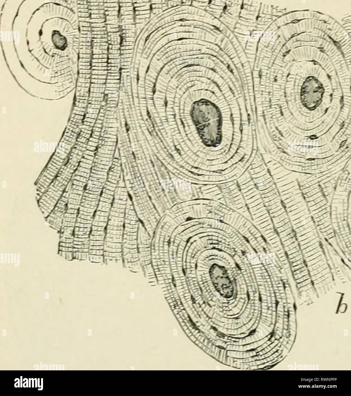
Compact bone is very different from the other tissues you have seen. Also called cortical bone, the compact variety usually features a haversian system, or cylindrical unit within the structure. In long bones, as you move from the outer cortical compact bone to the inner medullary cavity, the bone transitions to spongy bone. Transverse section of compact bone (metatarsal); Concentric layers of bone cells (osteocytes) and bone matrix surround the central canal. Compact bone is laid in such a manner that there are histological units seen in cross section. They conduct blood vessels, lymphatics, and nerves throughout the bone. Compact bone cross section courtesy: Because of its strength, the compact bone makes it possible for the bone to support weight. The osteocytes are arranged in concentric rings of bone matrix called lamellae (little plates), and their processes run in interconnecting canaliculi. Such roundish unit is called osteon. It is dense (because of calcified matrix) with tiny spaces known as lucanas. The compact bone is the main structure in the body for support, protection, and movement.
In the last decade, considerable technological improvements have been made to repair damaged bones and tissue, such as bone cross sections with implants for microscopic examinations. Compact bone is laid in such a manner that there are histological units seen in cross section. This is known as the periosteum. Concentric layers of bone cells (osteocytes) and bone matrix surround the central canal. They conduct blood vessels, lymphatics, and nerves throughout the bone.
Cross Section Of Compact Bone Flashcards Quizlet from quizlet.com Compact cross section human, ground bone, 162 x. The compact bone is the main structure in the body for support, protection, and movement. This photo shows a cross section through bone. Also called cortical bone, the compact variety usually features a haversian system, or cylindrical unit within the structure. When compact bone is studied, it is found to be made up of concentric circles called lamellae. Use colored pencils to draw and label the following structures as they appear using the 40x objective, or by looking at an image from the internet. Compact bone is laid in such a manner that there are histological units seen in cross section. These are abundant and characteristic of compact bone.
It consists of two layers;
Spongy bone is used for more active functions of the bones, including blood cell production and ion exchange. The osteon has blood vessels and bone cells, things vital for the survival of the bone. Such roundish unit is called osteon. Compact bone, makes up the dense material in a long section of a bone. The large dark spots are passages for blood vessels and nerves. There are two ways to study bone histology. The larger circular profiles are haversian canals and the smaller profiles are lacunae. Then, fill in the table below to describe each. Skull bone is a flat bone. The osteocytes are arranged in concentric rings of bone matrix called lamellae (little plates), and their processes run in interconnecting canaliculi. In the center of each osteon is the central canal, a space that houses blood vessels and nerves that supply bone. When compact bone is studied, it is found to be made up of concentric circles called lamellae. Compact bone is very different from the other tissues you have seen.
Start studying cross section of compact bone. However, compact bones also serve a function in storing and releasing calcium to the. It needs to be very strong as it supports your body and muscles as you walk, run, and move throughout the day. In this image the bar indicates the location of decalcified compact bone. Skull bone is a flat bone.
Elements Of Histology 1898 Elements Of Histology Elementsofhistol00klei Year 1898 72 Elemexts Of Histology Bones And In The Outer Layer Of Flat And Short Bones Its Lamell E Are Arranged As Concentric from c8.alamy.com To the left is muscle tissue, and to the right is bone marrow. Concentric layers of bone cells (osteocytes) and bone matrix surround the central canal. An outer 'fibrous layer' containing mainly fibroblasts, and an inner 'cambium layer' containing progenitor cells. Related posts of cross section of a long bone bone test anatomy and physiology. The compact bone is the main structure in the body for support, protection, and movement. In the center of each osteon is the central canal, a space that houses blood vessels and nerves that supply bone. Anatomy of a flat bone. Compact bone cross section courtesy:
Shop' die neuesten kollektionen bequem online bei surfdome jetzt.
The compact bone is made up of osteon. Compact bone is laid in such a manner that there are histological units seen in cross section. Note the holes in the eosinophilic matrix of the decalcified bone. Such roundish unit is called osteon. Their course follows the main axis of long bone. It needs to be very strong as it supports your body and muscles as you walk, run, and move throughout the day. The larger circular profiles are haversian canals and the smaller profiles are lacunae. Compact bone is very different from the other tissues you have seen. In long bones, as you move from the outer cortical compact bone to the inner medullary cavity, the bone transitions to spongy bone. Bone is hard and many of its functions depend on that characteristic hardness. These are abundant and characteristic of compact bone. Compact bone cross section courtesy: The osteocytes are arranged in concentric rings of bone matrix called lamellae (little plates), and their processes run in interconnecting canaliculi.
Also called cortical bone, the compact variety usually features a haversian system, or cylindrical unit within the structure cross section of a bone. Concentric layers of bone cells (osteocytes) and bone matrix surround the central canal.